A pregnancy is an unforgettable experience of a lovely journey as you go through a lot of exquisite and glorious experiences during pregnancy; one of which is watching a little life moving on a screen during your ultrasound. A pregnancy scan is not just a routine check-up; it’s often the first real glimpse into a new life.
The NT scan (Nuchal Translucency scan) is an ultrasound scan done during the first trimester of pregnancy as part of screening tests. This test helps in the detection of congenital abnormalities in the growing baby.
What is an NT Scan in Pregnancy?
It is a screening test, not a diagnostic test, as it just reports the level of risk of developing chromosomal abnormalities or congenital diseases in the growing baby.
It is an optional test and can be done along with other prenatal screening tests during the first trimester.
Generally, a baby has 46 chromosomes (23 pairs of chromosomes), which consist of two types of chromosomes — mainly 44 autosomal chromosomes and 2 sex chromosomes.
So, a developing baby boy has 46 chromosomes (22 pairs + XY chromosomes), and a developing baby girl has 46 chromosomes (22 pairs + XX chromosomes. Any imbalance of chromosomes can lead to abnormalities like Down syndrome, Patau syndrome, and Edward syndrome. In this test, high-frequency sound waves pass through the body and analyze the echoes that reflect back. These echoes are converted into electrical signals and processed to create real-time images of the developing baby.
During the NT scan, doctors look at the fluid-filled area at the back of the baby’s neck, known as the nuchal fold or nuchal translucency.
Generally, this scan is done up to the 14th week of pregnancy. The thickness of this fluid is measured at the back of the baby’s neck, which usually decreases as the gestation period progresses. Results are made on the basis of the thickness of the fluid-filled area.
As this is just a screening test, it doesn’t diagnose any condition; it only gives an idea about the risk of developing congenital diseases.
A Double Marker Test is usually done along with the NT scan to increase the accuracy of the results. Both tests are non-invasive and predictive in nature.
When This Scan Can Be Performed During Pregnancy
As this scan measures the amount and thickness of the fluid-filled area at the back of the neck of the growing baby, this fluid starts regressing after the 14th to 15th week of gestation. Therefore, it is recommended to perform the NT scan during the 11th to 14th week of gestation.
During this time, other screening or prenatal tests are also performed in the first trimester. The best period for the NT scan is between the 11th and 14th week of pregnancy.
Why It Is Important to Do NT Scan
NT scan is always important to be done during pregnancy as it helps in:
- Early detection of genetic conditions
An NT scan is performed during the first trimester.
It gives an idea about the risk of developing abnormalities at the early stage of pregnancy.
Therefore, better management can be provided if available, and it gives guidance about the need for diagnostic tests for confirmation. - Non-invasive test
It is just an ultrasound scan, which is non-invasive in nature; therefore, it doesn’t harm the baby or the expecting mother. - Guidance for further diagnostic testing
If the NT scan shows high fluid in the back of the neck of the developing baby, then the baby has a high risk of developing genetic abnormalities.
This scan helps to differentiate in which cases a diagnostic test is necessary and in which it is not necessary to perform. - Peace of Mind / Reassurance
This scan can provide assurance to the parents about their growing baby’s health status — whether the baby is developing normally or not, and if there are any genetic or congenital abnormalities. - Double Marker Test
As Double Marker Test is generally performed with the NT scan (ultrasound test), it gives better results as it improves the accuracy of the report.
Which One Must Undergo the NT Scan?
NT scan is recommended when:
- You are more than 35 years of age.
- There is any past or current history of genetic abnormalities.
- Abnormal ultrasound findings in prior scans.
But it’s important to know that the NT scan is a screening test.
It just tells about the developing structure — whether it is normal or not (congenital diseases) — and the risk of having genetic abnormalities.
Diagnostic tests are needed to confirm the condition or disease.
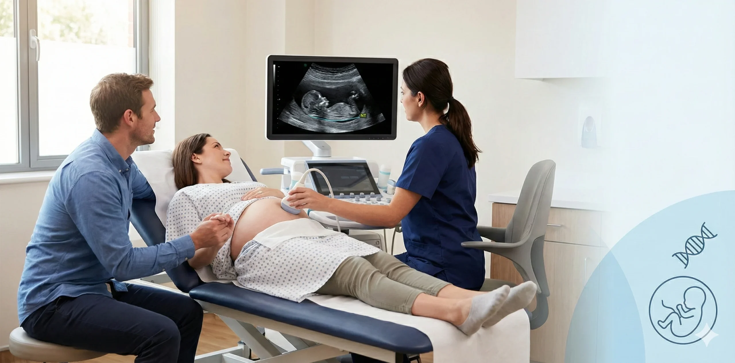
How It Is Done / Procedure of NT Scan
It is simply done as an ultrasound test. Typically, it is performed in every clinic or hospital setting and usually takes around 30 minutes to perform.
- The mother is asked to lie on an examination table, and a gel is applied to the abdomen area of the mother.
- The ultrasound probe is then moved over the abdomen.
In this process, high-frequency sound waves are transmitted into the body, and in return, echoes are produced, which result in the formation of a real-time image. - The amount and thickness of the nuchal translucency are measured and assessed by the doctor.
Besides this, the crown-rump length is also measured, which helps in knowing the gestational age. - These measurements are assessed by doctors along with the results of other screening tests, and then they calculate the risk of having chromosomal abnormalities in the growing baby.
Risk Associated with NT Scan
An NT scan is a non-invasive test performed during the 11th to 14th week of gestation. Therefore, it causes no harm to the growing baby or mother.
Results Interpretation of NT Scan
Results can be standard (normal) or abnormal. We will discuss normal/standard results.
Standard Results of NT Scan:
This means the thickness of Nuchal Translucency lies within the normal range, which is considered to be less than 3.5 mm for a baby at 11–14 weeks of gestational age.
Abnormal Results of NT Scan:
This means the thickness of Nuchal Translucency is greater than 3.5 mm, which increases the risk of developing structural defects in the growing baby. Therefore, to confirm any condition, a diagnostic test is required.
| NT Scan Result | Measurement (11–14 weeks) | Implication / Risk | Recommended Action |
| Normal | < 3.5 mm | Low risk of chromosomal abnormalities (e.g., Down syndrome, Edward syndrome, Patau syndrome) | Routine prenatal care; no additional diagnostic test required unless other risk factors exist |
| Abnormal / High Risk | > 3.5 mm | Increased risk of chromosomal abnormalities or structural defects | Consider diagnostic testing: NIPT, CVS (11–14 weeks), or Amniocentesis (after 15 weeks) |
Research Analysis:
A large population-based study in Ontario, Canada examined 414,268 singleton pregnancies to figure out if there’s a connection between NT thickness and chromosomal problems.
The study found that risk increases even when NT is below the commonly used 3.5 mm threshold.
Key Findings:
- Lowest Risk: NT < 2.0 mm (0.5% risk of chromosomal anomalies)
- Moderate NT (3.0–3.5 mm): 20-fold higher risk compared to NT < 2.0 mm (ARR 20.33; ARD 9.94%)
- Beyond Common Aneuploidies: Elevated risk remains for rarer chromosomal anomalies (ARR 4.97; ARD 1.40%)
Implication: Even NT measurements below 3.5 mm can indicate significant risk, suggesting the traditional threshold may need reconsideration. NT scan should be combined with other first-trimester tests like Double Marker Test or NIPT for more accurate risk assessment.
Reference:
Khalil A, et al. JAMA Netw Open. 2025
What Happens After Abnormal Findings
If your NT scan shows a reading greater than 3.5 mm of thickness, then it increases the chances of having chromosomal abnormalities. So, it’s just a screening test; to get confirmation, you need to undergo a diagnostic test. These include:
- Non-Invasive Prenatal Testing
This test analyzes the fetal DNA fragments in maternal blood. It can detect trisomies with 99% accuracy. - Chorionic Villus Sampling
Done during the 11th to 14th weeks of pregnancy. In this, a small sample of placental tissue is taken for genetic analysis. This test shows accuracy of more than 99%. - Amniocentesis
Done after the 15th week of pregnancy. In this test, a sample from the amniotic fluid is taken for genetic analysis. This also shows accuracy of more than 99%.
How to Prepare for NT Scan
- Non-Fasting Test: No fasting is needed before the NT scan. Eat normally unless advised by your doctor.
- Drink Water Beforehand: This helps in a fully dilated bladder, which results in a clearer image in early pregnancy.
- Drink Water: Try to drink 2–3 glasses of water about 40–60 minutes before the scan.
- Wear Comfortable Clothing: To easily assess your abdomen, loose clothing is helpful (like a tee & trousers).
- Expect a Trans-Abdominal Ultrasound: Most commonly, NT scans are done via the abdominal area of the body and rarely via a transvaginal scan.
Limitation of NT Scan
- Not Always Conclusive: Sometimes, NT is normal, but there are still chances of the presence of an abnormality, as the NT scan has a limited scope. (It may not detect all genetic or structural abnormalities).
- False +ve & False -ve
- False +ve can cause unnecessary stress and anxiety and lead to invasive tests.
- False -ve may lead to failure in screening for any chromosomal abnormality.
- Does Not Detect Neural Tube Defects: NT scan doesn’t assess conditions like spina bifida. That’s typically screened during the mid-pregnancy scan.
- Image Accuracy May Get Affected: Accuracy may be affected by many reasons, such as unfavourable fetal position, high maternal BMI, etc.
Case Study
Let’s understand this with a case study — a 33-year-old woman comes to the OPD block at 12 weeks of gestational age, conceived via in vitro fertilization. She undergoes prenatal screening tests. Her NT scan shows increased NT, rising up to 5.2 mm. On the basis of this NT scan result, her baby has a risk of developing chromosomal abnormalities.
After getting her screening test, she is advised to get genetic counselling from a counsellor. The counsellor recommended undergoing non-invasive prenatal testing, as this test doesn’t harm the developing baby or mother. All diagnostic tests are negative.
Outcomes: All her diagnostic tests are negative. She delivered a normal, healthy baby girl.
Frequently Asked Questions (FAQs)
Q1. Is NT Scan a mandatory test to go through in the 1st trimester?
Ans: No, it’s an optional screening test, which is recommended for every pregnant woman.
Q2. Can a normal NT scan rule out all abnormalities?
Ans: No, a normal NT scan doesn’t mean that your baby is 100 percent free from chromosomal abnormalities.
Q3. What is the role of the nasal bone in NT scan?
Ans: Absence or malformation of the nasal bone can be considered as another soft marker for the risk of having chromosomal abnormalities.
Q4. Is this test considered under insurance?
Ans: It depends on the healthcare system and the insurance provider. Many private plans cover it as part of prenatal care.
Q5. What if the baby’s position makes measurement difficult?
Ans: In this case, your sonographer will advise you to come later after some time, and most of the measurements become possible later.
Note from Femwise Health
Any extra chromosome (more than the usual 23 pairs of chromosomes) is considered an abnormal condition and results in different chromosomal abnormalities like Down syndrome, Patau syndrome, Edward syndrome, etc. Hope this blog helps you understand the details about the NT scan test and why it is necessary to go through this test.
Glossary Terms
- Nuchal Translucency: It is a fluid-filled space present at the back of the neck of the developing baby and measured in the early duration of pregnancy, i.e., during the 11th to 14th week of gestational age.
- Trisomy 21: Trisomy of the 21st chromosome is called Down syndrome, in which an extra copy of the 21st chromosome is formed.
- Trisomy 18: Trisomy of the 18th chromosome is called Edward syndrome, in which an extra copy of the 18th chromosome is formed.
- Trisomy 13: Trisomy of the 13th chromosome is called Patau syndrome, in which an extra copy of the 13th chromosome is formed.
- Beta hCG: A hormone produced only during the pregnancy phase in women. It is used as a marker in many pregnancy tests.
- CRL (Crown Rump Length): It’s the length of the baby from head to bottom, measured to determine the gestational age.
- Screening Test: This test only gives an estimate of the chances of having risks but never confirms any diagnosis.
- Diagnostic Test: This test gives confirmation of having any condition and is mostly done after the screening test.
- Gestational age – Age of the pregnancy from the last date of the menses.


